
Research
Our lab develops quantitative imaging technologies to study gene regulation from single molecules to intact tissues. Our research connects three scales: molecular dynamics, genome organization, and spatially resolved cell and tissue biology.
Our Working Hypothesis
We hypothesize that many genomic and epigenetic features are not primarily acting as simple switches for individual genes. Instead, they may fine-tune coordinated gene expression across many genes at the single-cell level.
By analogy, these regulatory interactions can act like the weights of a neural network, tuning relationships among genes and shaping collective expression states.
These effects can be subtle, yet collectively they can shape cell state, spatial gene-expression patterns, and tissue function. Detecting and testing such effects requires single-cell measurements with low noise, high dynamic range, and sufficient multiplexing to resolve many molecular variables across molecular, genomic, cellular, and tissue scales.
The precision of the measurement is itself part of the experiment.
Molecular Dynamics
We develop approaches for tracking and labeling individual proteins in living cells and whole organisms, including residence-time-resolved transcription factor imaging (Chen et al., Cell 2014) and stochastic protein labeling for long-term single-molecule imaging in vivo (Liu et al., PNAS 2018).
These tools have revealed how transcription factors search the genome, how molecular cargo is transported and sorted in neurons, and how disease-associated protein aggregates disrupt molecular dynamics and gene regulation.
Genome Organization
We invented 3D ATAC-PALM, a super-resolution imaging method that combines the Assay for Transposase-Accessible Chromatin with lattice light-sheet PALM microscopy to visualize nanoscale genome organization in single cells (Xie & Dong et al., Nature Methods 2020).
Using 3D ATAC-PALM with genetic perturbations, we discovered that cohesin compartmentalizes the accessible genome and regulates patterns of gene co-expression in single cells, even when changes in average gene expression are modest (Xie & Dong et al., Nature Genetics 2022; Dong et al., Nature Genetics 2024).
These findings suggest that genome organization can regulate relationships among genes in ways that are not apparent from population-average expression measurements.
Cells & Tissues
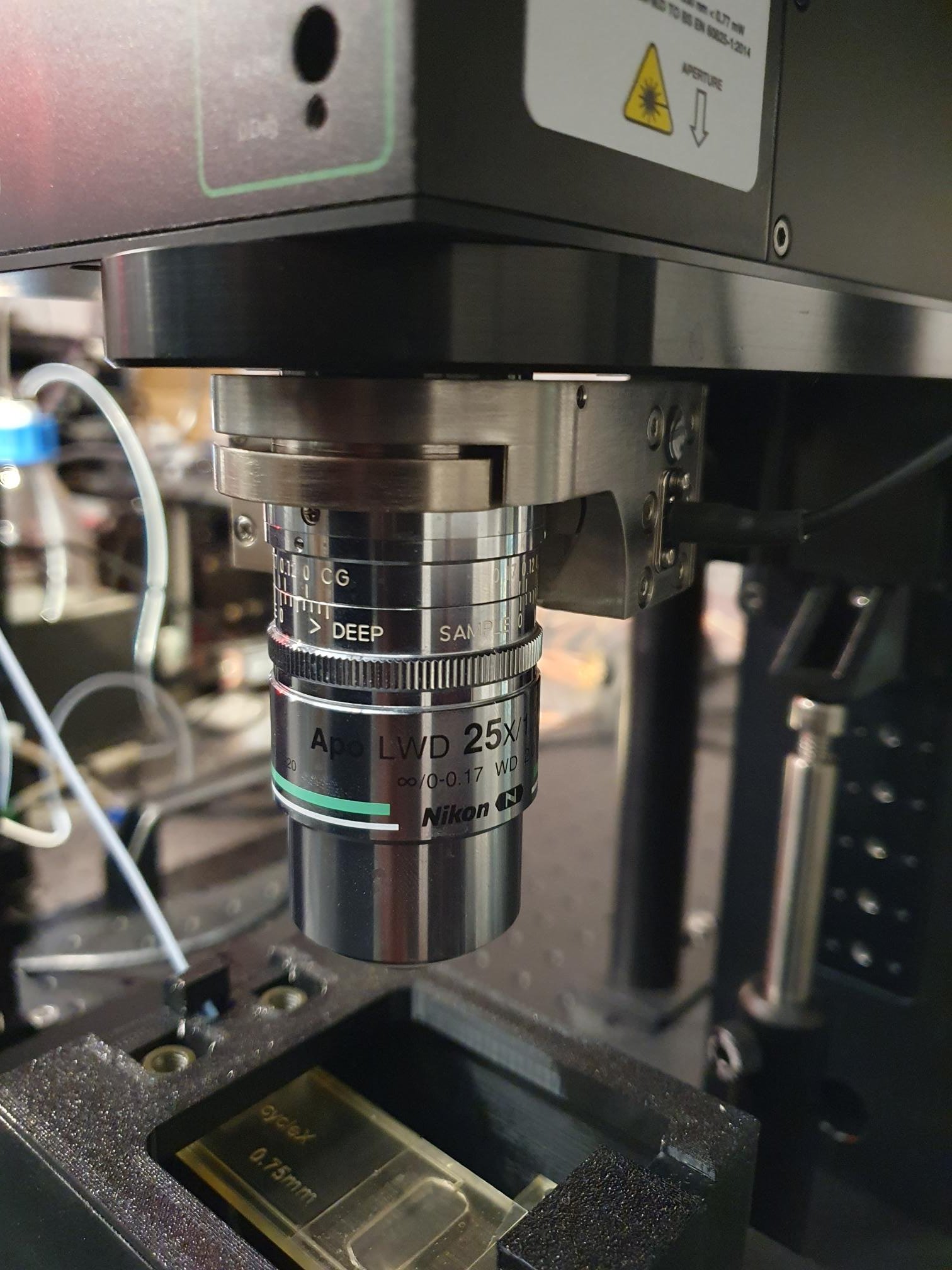
To understand how molecular and regulatory states are organized across intact tissues, we developed cycleHCR, a deep-tissue spatial transcriptomics and proteomics platform that images hundreds of RNA and protein targets with subcellular resolution in tissue volumes greater than 300 µm (Gandin & Kim et al., Science 2025).
cycleHCR integrates molecular chemistry, automated fluidics, high-resolution imaging, and computational analysis to measure molecular states while preserving their native cellular and tissue context.
We are using these approaches to study development, brain organization, cell-state variation, and cell-type-specific molecular architecture.
Connecting the Scales
Together, these approaches connect molecular dynamics, genome organization, cellular states, and tissue context. By combining quantitative imaging with perturbation experiments and computation, we aim to understand how dynamic molecular organization gives rise to cell state and tissue-level physiological function.

Deep-tissue multiplexed RNA (254) imaging | Learn More →
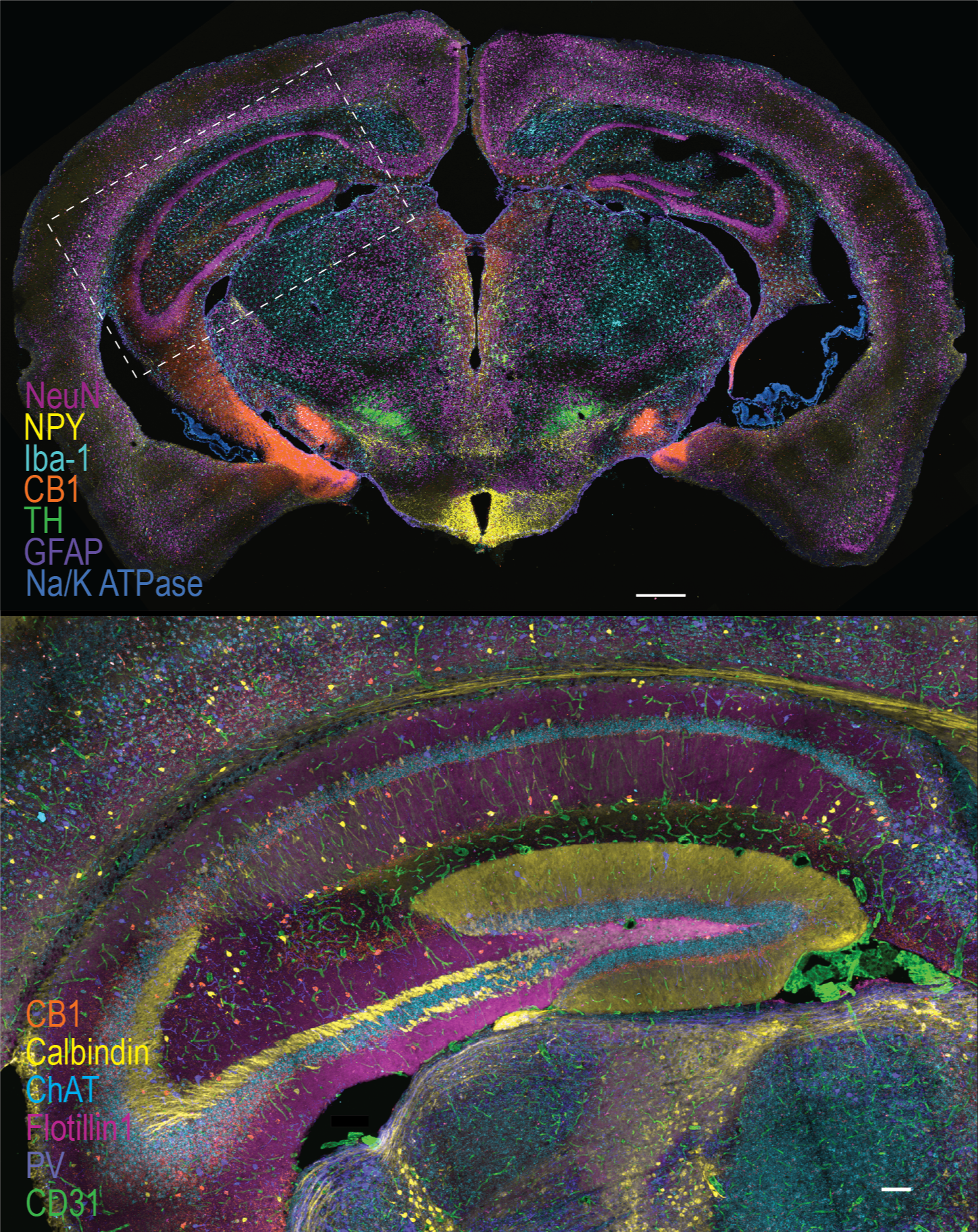
Multiplexed Protein (46) + RNA (79) imaging in whole brain slices | Learn More →